TCIA-DEMO-001
CT CHEST WITHOUT CONTRAST · AXIAL 1.25 mm · 206 images · STANDARD kernel
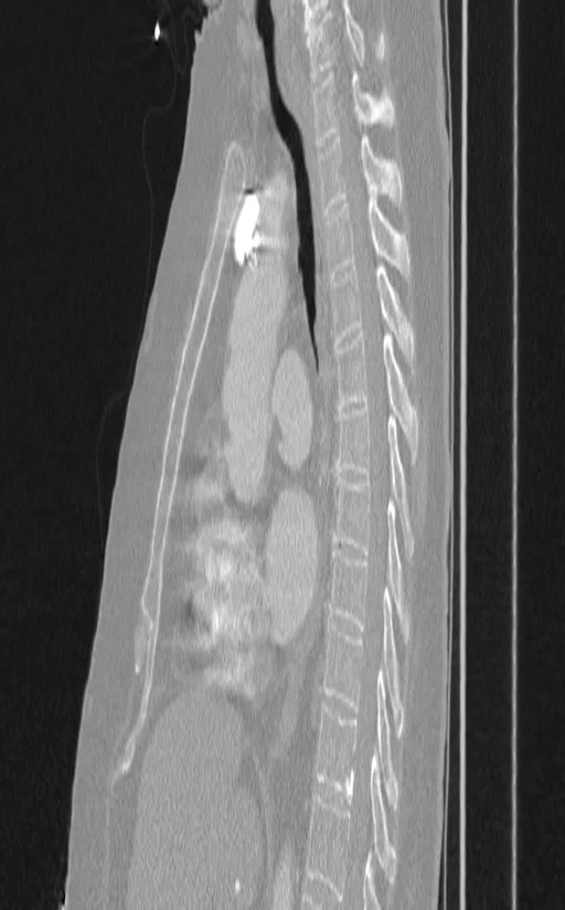
LIDC-IDRI — The Cancer Imaging Archive (TCIA)
De-identified at source. Patient name removed, study date shifted.
Licensed CC BY 3.0 · DOI 10.7937/K9/TCIA.2015.LO9QL9SX
Precision. The slices ship as 8-bit samples across a fixed −1200…+800 HU window and the encoder is lossy, so density is quantised to roughly 8 HU with further error on top. Window and level run against real Hounsfield units recovered from those samples, but every readout is rounded to 10 HU and marked ≈: it shows the scale is real, not that the number is a measurement. A window above +800 HU clamps, so bone presets are limited.
The authors acknowledge the National Cancer Institute and the Foundation for the National Institutes of Health, and their critical role in the creation of the free publicly available LIDC/IDRI Database used in this study.